Categories
Blogs

IOUS guided liver surgery

IOUS guided liver surgery

Pushing the Limits of cancer surgery - challenging dogmas and treading the thin line

As young and eager type A surgical residents, we were all told this story to rein in our horses. Most surgical oncologists must have heard some or the other version of it. Here’s the version I heard – there was this young surgical registrar who was performing a total esophagectomy and realised that the oesophageal lesion was locally advanced and probably inoperable. He called in his lecturer who after a long battle managed to remove the tumour and left the OT with a swagger, believing he had done a good job. The next time, this lecturer was in trouble and the Assistant professor had to step in to rescue him and remove the tumour.

This same incident repeated with the Assistant professor and the HOD had to step in. The resident felt that he had a long way to go, which he did, but at the end of one year – all the patients were dead from a recurrence.

 Moral of the story:

  1. The lighter version which, as a resident I liked was that “the Resident is always right about inoperability”.

  2. The real lesson that was intended for the residents was –  “There is no role of surgical bravado in the treatment of cancer”. This was perhaps taught to us so that we would follow protocols and ensure standardized outcomes for patients in a teaching institute.

But as you grow older and wiser, you realize that this philosophy is regressive and deters evolution of surgical techniques. Take the case of Liver surgery for colorectal liver metastases (CRLM). In the 1980s and 90s, there were stringent selection criteria for consideration for a liver resection – <4 lesions, confined to one lobe, <5cm size and absence of extra-hepatic disease. But this was in the era before doublet chemotherapies like FOLFOX or FOLFIRI, which we now know are far superior to then existent chemotherapy options. Then came the era of biological therapy, which in combination with optimal chemotherapy gives responses that even converted initially inoperable CRLM to operable in 20% cases. And this changed the landscape of surgery for CRLM.

But was it only the chemotherapy and biological therapy that made the difference? Had it not been for curious and adventurous surgeons who wanted to push the envelope this wouldn’t have been possible. We now have surgical strategies like 2-stage liver resection in combination with portal vein embolisation, liver-first approach, ALPPS, intra-operative ultrasound (IOUS) guided liver surgery, and indeed liver transplantation for inoperable liver metastases. And all ancillary modalities have developed around these adventurous surgeons. Which is why we now have better and safer anaesthesia, highly accomplished interventional radiologists, better energy devices, sophisticated intra-operative imaging techniques and better post-operative care units.

Of course, these surgeons who think differently and challenge dogmas need the support of their colleagues, medical oncologists, interventional and conventional radiologists; and indeed their institutions to do the research, develop protocols, get ethics committee approval, perform these challenging surgeries, and ensure good outcomes. Over the last 3 decades, treatment of CRLM has become a sub-specialty in itself and more and more studies have pointed out the importance of concentrating these cases in highly specialized units to ensure better outcomes.

Our own experience has shown a trend towards shorter hospital stay and better post-operative outcomes with the adoption of IOUS and parenchyma-sparing liver surgery. In this treatment strategy, we use the latest devices in intra-operative imaging (HITACHI ALOKA ARRIETA 70 with the micro-convex echo probe for open surgeries and 4-directional Laparoscopic probe for laparoscopic surgeries) to mark out the anatomical landmarks and the exact locations of the liver lesions in relation to these structures. Based on pre-operative imaging and volumetric studies along with intra-operative IOUS findings, a 3D map of the liver to be resected can be envisaged and a highly precise resection is performed to remove all the CRLM while preserving as much of the normal liver parenchyma as possible along with its portal supply and venous drainage. Often, the challenge in extensive liver metastases is the preservation of adequate venous drainage; IOUS guided flow analysis helps in the detection of collateral communications among hepatic veins that helps tailor the hepatic  resections beyond the standard segmentectomies, sectionectomies and hemihepatectomies.
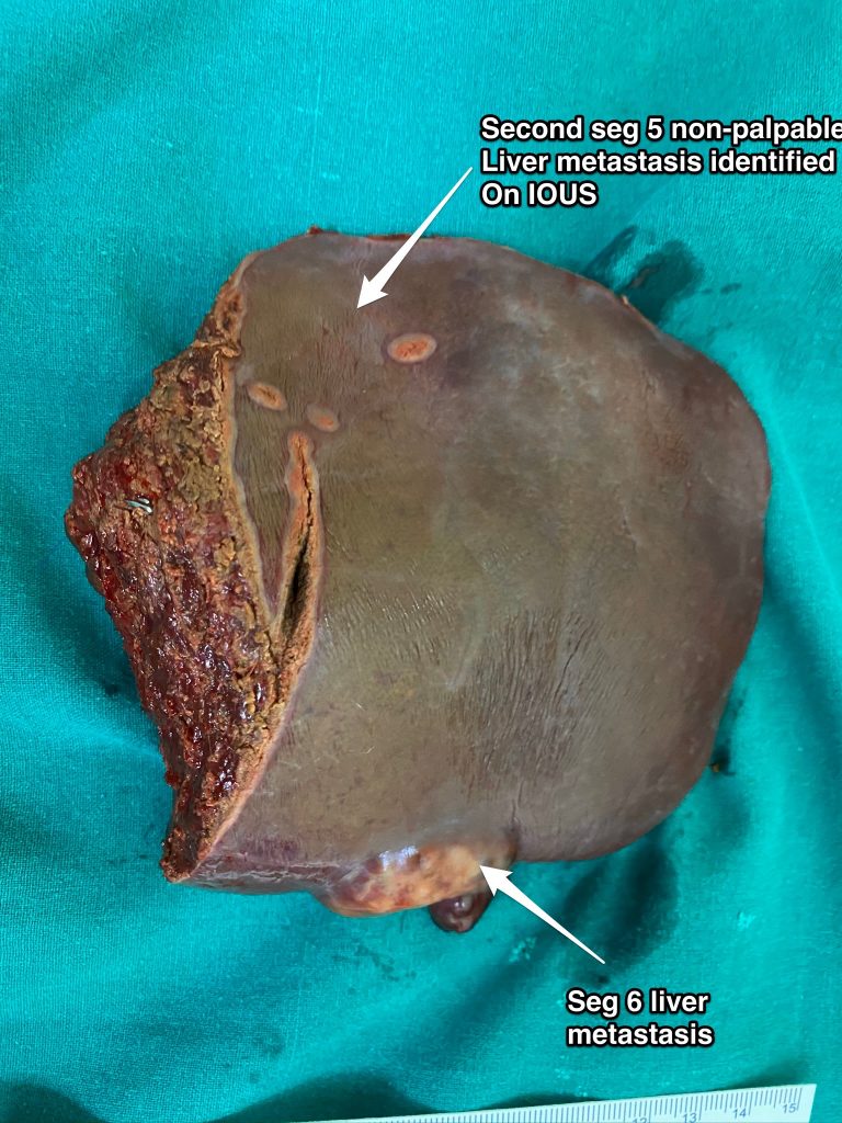
Fig 1 A

Fig 1 B

Fig. 1A and 1B – Lesion in seg 6 and another non-palpable lesion in segment 5. In the absence of IOUS, it would involve a formal set 5 resection but with IOUS, a contiguous resection can be performed with adequate margins without sacrificing additional normal liver parenchyma

Fig 2A – IOUS identifying 3 lesions straddling segments 7,8 above the plane of the hepatic veins

Fig 2B – Resection performed under IOUS guidance to resect segments 7 and 8 above the plane of the hepatic veins to encompass the 3 liver metastases. An additional small liver metastasis identified in segment 6 was removed separately

Fig 2C – Final specimens
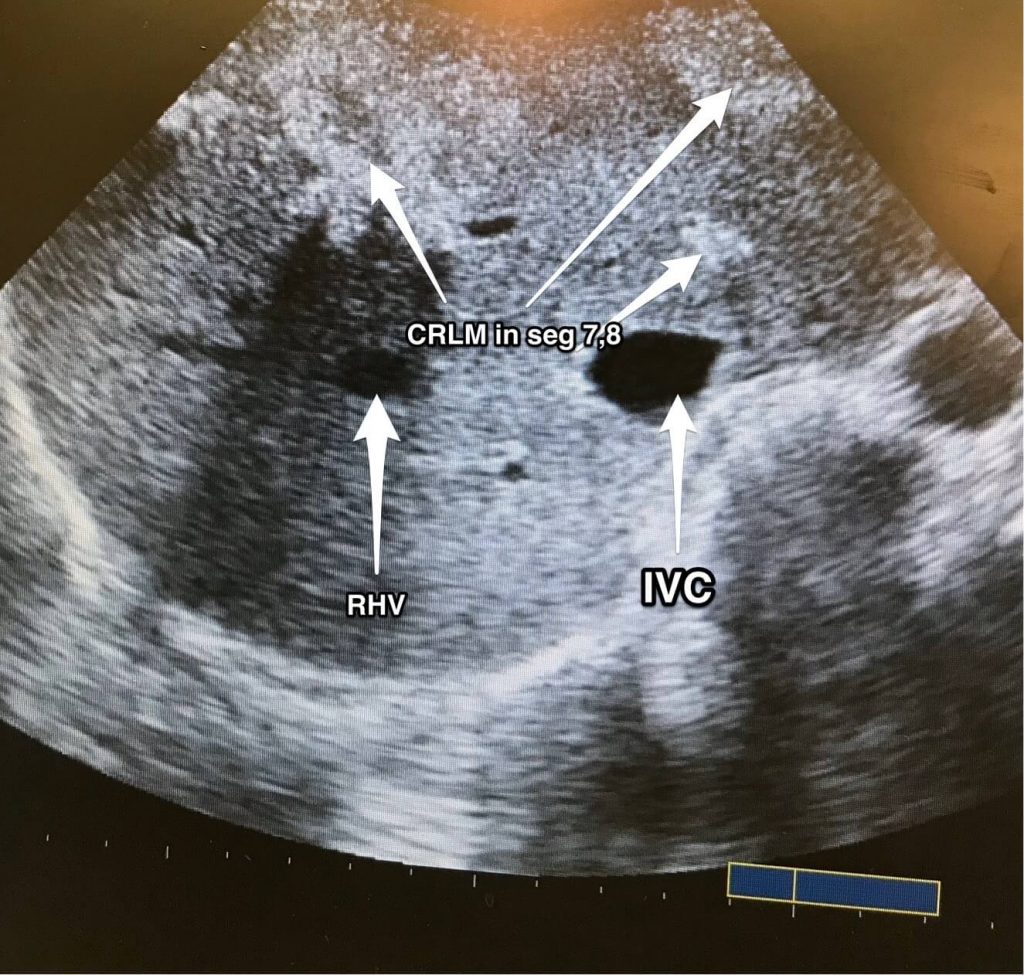
Fig 2 A

Fig 2 B

Fig 2 C
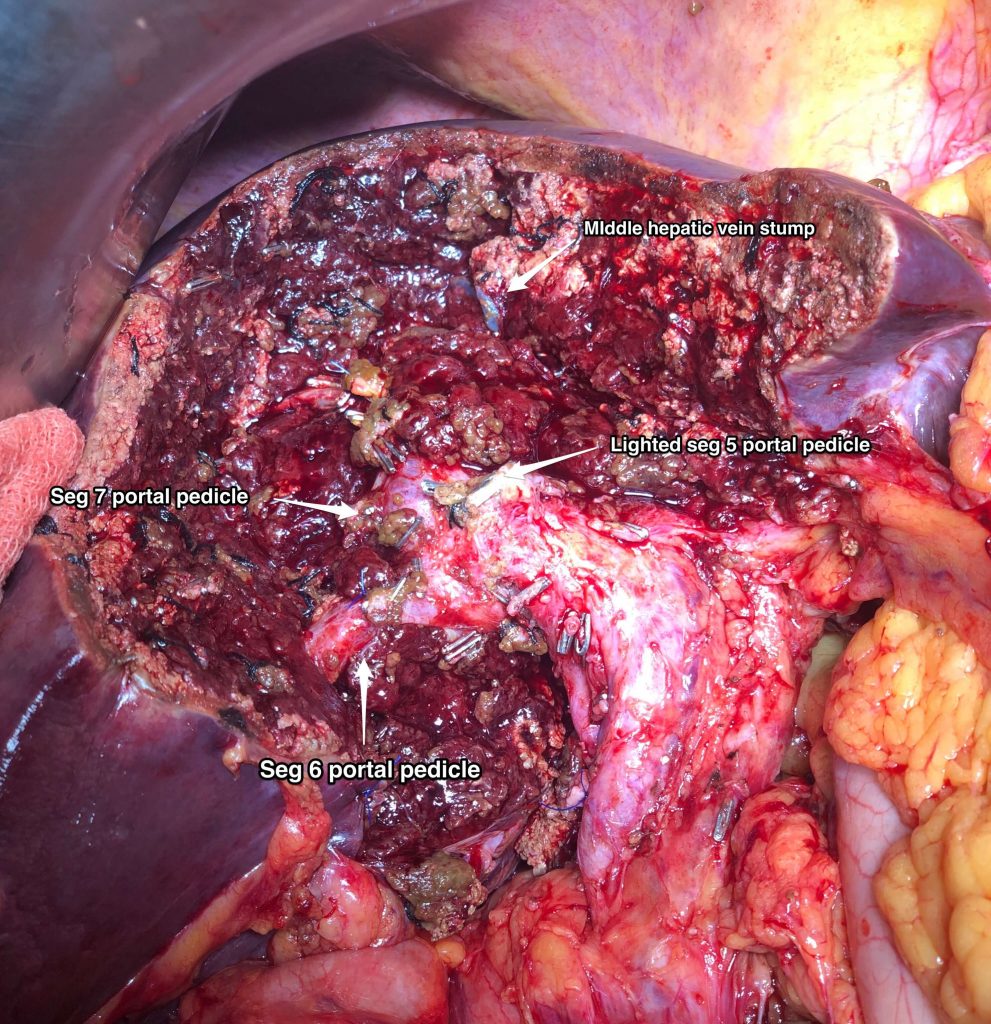
Fig 3 - A precise resection of seg 4B, 5, 8p done under IOUS guidance to resect large CRLM in close relation to the right portal pedicle

Categories
Blogs

Appendix Tumors

Appendix Tumors

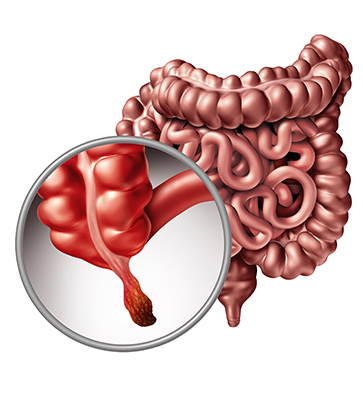
The appendix is a pouch-like tube that is attached to the cecum (the first section of the large intestine or colon). The appendix averages 10 centimeters (cm) in length and is considered part of the gastrointestinal (GI) tract. Generally thought to have no significant function in the body, the appendix may be a part of the lymphatic, exocrine, or endocrine systems.

Appendix cancer occurs when cells in the appendix become abnormal and multiply without control. These cells form a growth of tissue, called a tumor. A tumor can be benign (noncancerous) or malignant (cancerous, meaning it can spread to other parts of the body). Another name for this type of cancer is appendiceal cancer.

Types of Appendix Tumors

Carcinoid Tumor
A carcinoid tumor starts in the hormone-producing cells that are normally present in small amounts in almost every organ in the body. A carcinoid tumor arises primarily in either the GI tract or lungs, but it also may occur in the pancreas, a man’s testicles, or a woman’s ovaries. An appendix carcinoid tumor most often occurs at the tip of the appendix. Approximately 66% of all appendix tumors are carcinoid tumors. This type of cancer usually causes no symptoms until it has spread to other organs and often goes unnoticed until it is found during an examination or procedure performed for another reason. An appendix carcinoid tumor that remains confined to the area where it started has a high chance of successful treatment with surgery.
Colonic-Type Adenocarcinoma
Colonic-type adenocarcinoma accounts for about 10% of appendix tumors and usually occurs at the base of the appendix. This type of tumor looks and behaves like the most common type of colorectal cancer. It often goes unnoticed, and diagnosis is frequently made during or after surgery for appendicitis (inflammation of the appendix that can cause abdominal pain or swelling, loss of appetite, nausea, vomiting, constipation or diarrhea, inability to pass gas, or a low fever that begins after other symptoms).
Signet-Ring Cell Adenocarcinoma
Signet-ring cell adenocarcinoma (so called because, under the microscope, the cell looks like it has a signet ring inside it) is very rare and considered to be more aggressive and more difficult to treat than other types of adenocarcinomas. This type of tumor usually occurs in the stomach or colon, and it can cause appendicitis when it develops in the appendix.
Paraganglioma
Paraganglioma is a rare tumor that develops from cells of the paraganglia, a collection of cells that come from nerve tissue that persist in small deposits after fetal (pre-birth) development, and is found near the adrenal glands and some blood vessels and nerves. This type of tumor is usually considered benign and is often successfully treated with the complete surgical removal of the tumor. Paraganglioma is very rare outside of the head and neck region.
Mucinous Cystadenocarcinoma
Mucinous cystadenocarcinoma is the most common non-carcinoid appendix tumor and accounts for about 20% of appendix cancer cases. This type of tumor produces a jelly-like substance called mucin that can fill the abdominal cavity and can cause abdominal pain, bloating, and changes in bowel function if the tumor breaks through the appendix or grows in the abdomen. This is called as Pseudomyxoma Peritonei or “jelly belly”.
Appendiceal Tumors
Appendiceal tumors are often misdiagnosed as appendicitis. Alternatively, they may burst with very little symptoms and spread within the abdominal cavity to give rise to pseudomyxoma peritonei or peritoneal carcinomatosis. Fortunately, now we can treat these conditions using Cytoreductive Surgery and Hyperthermic Intra-peritoneal chemotherapy (HIPEC). Dr. Sanket Mehta has pioneered this form of treatment in the country. He has already performed numerous such surgeries with good results.
Categories
Medreads

Gastrointestinal & Colon Cancers

Gastrointestinal & Colon Cancers

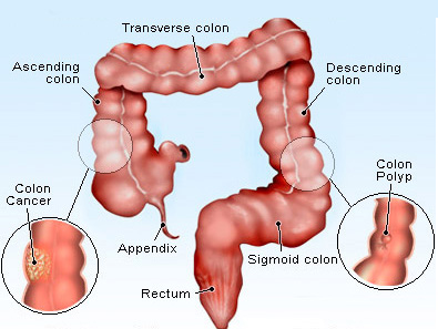
The colon is the part of the digestive system where the waste material is stored. The rectum is the end of the colon adjacent to the anus. Together, they form a long, muscular tube called the large intestine (also known as the large bowel). Tumors of the colon and rectum are growths arising from the inner wall of the large intestine. Benign tumors of the large intestine are called polyps. Malignant tumors of the large intestine are called cancers. Benign polyps do not invade nearby tissue or spread to other parts of the body. Benign polyps can be easily removed during colonoscopy and are not life-threatening. If benign polyps are not removed from the large intestine, they can become malignant (cancerous) over time. Most of the cancers of the large intestine are believed to have developed from polyps. Cancer of the colon and rectum (also referred to as colorectal cancer) can invade and damage adjacent tissues and organs. Cancer cells can also break away and spread to other parts of the body (such as liver and lung) where new tumors form. The spread of colon cancer to distant organs is called metastasis of the colon cancer. Once metastasis has occurred in colorectal cancer, a complete cure of the cancer is unlikely.

What are the causes of Colon Cancer?
Doctors are certain that colorectal cancer is not contagious (a person cannot catch the disease from a cancer patient). Some people are more likely to develop colorectal cancer than others. Factors that increase a person’s risk of colorectal cancer include high fat intake, a family history of colorectal cancer and polyps, the presence of polyps in the large intestine, and chronic ulcerative colitis.
Diet and Colon Cancer?
Diets high in fat are believed to predispose humans to colorectal cancer. In countries with high colorectal cancer rates, the fat intake by the population is much higher than in countries with low cancer rates. It is believed that the breakdown products of fat metabolism lead to the formation of cancer-causing chemicals (carcinogens). Diets high in vegetables and high-fiber foods such as whole-grain breads and cereals may rid the bowel of these carcinogens and help reduce the risk of cancer.
Colon Polyps and Colon Cancer?
Doctors believe that most colon cancers develop in colon polyps. Therefore, removing benign colon polyps can prevent colorectal cancer. Colon polyps develop when chromosome damage occurs in cells of the inner lining of the colon. Chromosomes contain genetic information inherited from each parent. Normally, healthy chromosomes control the growth of cells in an orderly manner. When chromosomes are damaged, cell growth becomes uncontrolled, resulting in masses of extra tissue (polyps). Colon polyps are initially benign. Over years, benign colon polyps can acquire additional chromosome damage to become cancerous.
Categories
Medreads

Peritoneal Mesothelioma

Peritoneal Mesothelioma

Malignant mesothelioma is a rare form of cancer that affects the thin cell wall lining of the body’s internal organs and structures. This lining is known as the mesothelium.

It is of three varieties:

  1. Pleural Mesothelioma (covering of the lungs)

  2. Peritoneal Mesothelioma (covering of abdomen)

  3. Pericardial Mesothelioma ( covering of heart)


Risks Factors:

Asbestos exposure is a known risk factor for development of malignant mesothelioma. Asbestos is a natural, yet toxic, mineral that was frequently used across a wide variety of industries. Microscopic asbestos fibers enter the body via the lungs or by means of ingestion. Once inside the human body, the durable asbestos fibers are unable to be broken down or expelled, resulting in a harmful inflammation and scarring of the mesothelium. This scarring lays the groundwork for malignant mesothelioma.

No other genetic, dietary or geographic factors or variation have been reported.

Signs and Symptoms:

Trouble breathing, pain under the rib cage, distension of abdomen due to ascitis, lumps in the abdomen, unexplained weight loss. The symptoms depend on the extent of involvement. Nowadays, a common mode of diagnosis is incidental finding on laparoscopy for infertility or other procedures.

How is it diagnosed?

  1. Imaging Scan: CT Scan, MRI, PET CT usually picks up the disease.

  2. Biopsy: Cytology cannot diagnose a mesothelioma. A biopsy is essential and the goal standard for the diagnosis of mesothelioma. A staging laparoscopy is ideal since it helps get adequate biopsy sample and assess the extent of peritoneal disease.

  3. Blood Tests: Tumor marker levels like Ca 125, Ca 19-9 and CEA are advisable but not diagnostic.

Categories
Medreads

Ovarian Cancer

Ovarian Cancer

Experts do not know exactly what causes ovarian cancer. But they do know that DNA changes play a role in many cancers.Some women are more likely than others to get this rare cancer. Women who are past menopause or who have never been pregnant are more likely to get ovarian cancer.
What are the Symptoms?
In some cases, ovarian cancer may not cause early symptoms. But most women do have symptoms, even in early-stage ovarian cancer. These symptoms include recent, frequent bloating; pain in the belly or pelvis; difficulty eating or feeling full quickly; or urinary problems, such as an urgent need to urinate or urinating more often than usual. Other symptoms that women with ovarian cancer may have include fatigue, indigestion, back pain, pain with intercourse, constipation, and changes in their menstrual cycles. But these symptoms are also common in women who don’t have ovarian cancer.
How is it Treated?
Surgery is the main treatment. The doctor will remove any tumors that he or she can see. This usually means taking out one or both ovaries. It may also mean taking out the fallopian tubes and uterus. After surgery, most women have several months of chemotherapy, which means taking drugs that kill cancer cells. This cancer often comes back after treatment. So you will need regular checkups for the rest of your life. If your cancer does come back, treatment may help you feel better and live longer.
Treatment of Overian Cancer
The treatment of ovarian cancer is based on the stage of the disease, which is a reflection of the extent of spread of the cancer to other parts of the body. Staging is performed by the surgeon when the ovarian cancer is removed. During the surgical procedure the surgeon will obtain small pieces of tissue (biopsies) from various sites in the abdominal cavity. During this procedure, depending on the stage (extent) of the disease, the surgeon will either remove just the ovary and fallopian tube or will remove both the ovaries, fallopian tubes and uterus. In addition, the surgeon will attempt to remove all visible cancer.
Overian Cancer is staged as follows:
Stage I cancer is confined to one or both ovaries. The cancer is Stage II if either one or both of the ovaries is involved and has spread to the uterus and/or the fallopian tubes or other sites in the pelvis. The cancer is Stage III cancer if one or both of the ovaries is involved and has spread to lymph nodes or other sites outside of the pelvis but is still within the abdominal cavity, such as the surface of the intestine or liver. The cancer is Stage IV cancer if one or both ovaries is involved and has spread outside the abdomen or has spread to the inside of the liver.
Treatment Options:
There are basically three forms of treatment of ovarian cancer. The primary one is surgery at which time the cancer is removed from the ovary and from all the other sites. Chemotherapy is the second important modality. This form of treatment uses drugs to kill the cancer cells. The other modality is radiation treatment, which is used in rare instances. It utilizes high energy x-rays to kill cancer cells. Surgical treatment is the single most important modality determining the outcome of ovarian cancer, besides its inherent biological behavior. It is best performed by a specialist oncosurgeon who has been specially trained in the diagnosis and management of gynecologic malignancy. The treatment of ovarian cancer depends on the stage of the disease, the histologic cell type, and the patient’s age and overall condition. The histologic cell type and the extent of disease based on the biopsies performed by the gynecologic oncologist during surgery (staging), are determined by the pathologist who analyzes tissues with a microscope.
Recurrent Overian Epithelial Cancer:
Detection of Recurrent Disease : Small tumors generally respond better to treatment, therefore early detection of recurrence may be useful. However it is important to consider that the benefits of early introduction of salvage chemotherapy are limited and may intrude upon the patient’s symptom and treatment-free survival. Use of frequent clinical follow-up can detect treatment failure earlier. Follow-up includes bimanual pelvic examination, serial measurement of CA125 or another tumor marker, reassessment or second-look laparotomy and occasionally one or more imaging studies. However, recurrent cancer has a large spectrum of behavior making it relatively difficult to diagnose a relapse and determine the aggressiveness of the tumor.
Second-Look Surgery
The use of second-look surgery can help diagnose and manage ovarian cancer. The typical indication for which a Second-look is performed at our center is when an incomplete Cytoreduction has been performed at the first instance (usually by an inexperienced surgeon). Another rare instance is when an incomplete Cytoreduction was performed due to the poor general condition of the patient at the time of the first surgery, which has significantly improved after the adjuvant intra-venous chemotherapy. There is a recently published multi-centric studyref that strongly supports the role of HIPEC in this situation with more than doubling of the survival compared to historical standards. We promote the use of HIPEC in this situation and have had encouraging results for our patients.
Treatment of Recurrent Cancer:
Patients who develop recurrent cancer despite surgery and primary chemotherapy, and will be given salvage chemotherapy, may be placed into one of three groups (A-C):
Group A: are patients resistant to primary therapy and have shown tumor growth during treatment. This persisting tumor is considered to be refractory i.e. have absolute platinum-resistance. Secondary non-cross resistant chemotherapies or biological therapies should be considered.
Group B: are patients who respond well to initial chemotherapy, but develop recurrent cancer within months after the end of primary care. This group with relatively platinum resistant tumor has an intermediate prognosis.
Group C: are patients who showed a good response to primary chemotherapy, and did not develop recurrent cancer for more than 6 months after the end of primary treatment. This group with platinum-sensitive tumor shows the best responses to re-treatment with a platinum-containing regimen. The probability of response to salvage chemotherapy is also markedly dependent upon the number of preceding chemotherapy regimens, such that third and fourth line chemotherapies are of limited benefit. However, unique patients responding to multiple retreatments with even the same regimen of chemotherapy are sometimes observed. Tumor burden, as assessed by the size of the largest lesion and the number of disease sites and histology (serous having the best outcome) are also independent predictors of response to salvage chemotherapy.
CRS and HIPEC in Recurrent Ovarian Cancers:
Selected patients with recurrent ovarian cancers can have a remarkable response with a well-performed CRS, especially if combined with HIPEC. Recent studiesref have shown that the survival can almost equal that of patients with primary stage III disease, provided a complete CRS (removal of all visible disease) can be achieved and combined with HIPEC. Usually such a surgery needs to be undertaken after some cycles of chemotherapy to ensure that all disease is resected. It is of paramount importance that a HIPEC expert undertakes this surgery to deliver optimal results. We have been regularly performing this surgery for recurrent ovarian cancers with encouraging outcomes.
Drug Resistance:
The likelihood a patient will respond to salvage chemotherapy correlates with, for the most part, the cancer’s degree of platinum drug resistance The Gynecological Oncology Group (GOG) defines platinum resistance as meeting any of the criteria listed below: Disease progression while on a first-line platinum-based regimen Tumor progression within 6 months of completion of platinum-based therapy Persistent clinically measurable disease with best response as stable disease at the completion of planned first-line therapy Persistent clinically measurable disease with best response as stable disease with rising CA 125 while receiving first-line non-protocol therapy. Rising CA 125 levels must be documented with two examinations where the last result being greater than or equal to 100. The most commonly used indicator of resistance is the period of time between the end of primary chemotherapy and relapse: the longer this length of time, the better the chances of responding to salvage chemotherapy. Cancer that relapses after primary treatment using a single platin analog will have less resistance to treatment than cancer that relapses after primary treatment using multi-agent treatment Generally, about 25%, 33%, and 60% respond to salvage treatment when their time between last chemotherapy and relapse is 6-12 months, 12-24 months, and greater than 24 months respectively. An integrated team approach between the surgeon and the medical oncologist is necessary to deliver good results in these complex situations. In rare instances, CRS and HIPEC can be performed even in drug-resistant recurrent ovarian cancers with encouraging results. However, these patients are selected after a thorough evaluation and counseling as not all patients will benefit from this treatment.
Categories
Medreads

Rectum Cancer

Rectum Cancer

Although colon cancer and rectal cancer share many features, there are important differences between these two diseases including, especially, the tendency for rectal cancer — but not colon cancer — to recur locally. Local recurrence of rectal cancer is common after standard surgery and is often catastrophic. It is difficult to cure, and the associated symptoms are debilitating. Accordingly, preventing local recurrence is one of the main treatment goals with rectal cancer.

The prognosis (outlook) with rectal cancer is clearly related to the degree of penetration of the tumor through the bowel wall and the presence or absence of lymph node involvement. These two characteristics form the basis for all staging systems developed for this disease.


The standard surgical procedure is called total mesorectal excision (TME) . Preoperative chemoradiotherapy has been found to reduce the risk of local recurrence and to cause fewer long-term toxic effects than if the chemoradiotherapy is given postoperatively. Broadly, at five years, the overall survival among patients with locally advanced rectal cancer, irrespective of whether they have had preoperative or postoperative chemoradiotherapy, is about 75%.

The treatment options for rectal cancer depend on several factors and the decision for each case is often an individualized decision for that particular patient. Certain guidelines are quite clear and if the patient falls within those criteria, the treatment is standard.

For Example Stomach Cancer

In case of early rectal cancer where the tumor is confined to the wall of the rectum and does not extend to the surrounding fat or mesorectum with no lymph node involvement, the treatment is surgery. Often this surgery will be performed laparoscopically or occassionally even through the anal canal (Transanal endoscopic surgery).
In cases where the carcinoma is very close to the anal sphincter (the muscle controlling defecation) and/or is large in size with lymph node spread or spread into the mesorectum (the fat surrounding the rectum), the best treatment option is to go for neo-adjuvant long-course chemo-radiotherapy (which is a combination of radiotherapy for 5 weeks along with oral chemotherapy) followed by a surgery after 6 weeks of completion of radiotherapy. The advantages of performing radiation prior to surgery are:

  • Increased rate of sphincter saving surgeries: This form of radiotherapy significantly reduces the bulk of the tumor and its extent of spread making a sphincter-saving surgery possible in several cases where otherwise an abdominoperineal resection with a permanent colostomy would be required.
  • Decreased local recurrence rate:Giving chemoradiation prior to surgery decreases the local recurrence rate of the cancer compared to the older approach of surgery followed by radiotherapy
  • Lesser long-term side-effects:Since the part being irradiated is removed later during surgery, the patient suffers very few long-term side-effects, especially with the more recent methods of delivery of radiotherapy.

The decrease in the tumor size also means that majority of these tumors can still be operated by laparoscopic techniques.

In certain circumstances a neo-adjuvant short-course radiotherapy may give an equivalent effect as a long-course radiotherapy. However, these cases have to carefully selected and individualized so as to maintain the efficacy of the treatment.

In cases where the cancer has already spread (for example to distant nodes or the liver or the lungs) but both the primary tumor and the metastatic site are operable, the surgery can still be performed with excellent outcomes. However, the surgery, chemotherapy and the radiotherapy have to be accurately timed to get the optimum outcome. In most such cases, the radiotherapy used may be short-course radiotherapy to decrease the time that the patient is off chemotherapy. Dr. Sanket Mehta regularly performs rectal cancer surgery laparoscopically. He specializes in the treatment of loco-regionally advanced and operable metastatic rectal cancer. He has performed several major synchronous resections of the rectum with liver metastases or peritoneal metastases, along with CRS and HIPEC in case of pertitoneal disease

Categories
Medreads

Pancreatic Cancer

Pancreatic Cancers

Pancreatic cancers are often aggressive cancers and the treatment depends on the extent of spread of the cancer and overall health of the patient. Your doctor will advice you the exact treatment plan after a thorough evaluation and adequate investigations which may include a CT scan, MRCP and even an endoscopic evaluation.

The main stay in the treatment of pancreatic cancer is surgery. Though the surgeries take long hours and involve complex procedures, with advances in anaesthesia, medical and intensive care, and better equipment; these surgeries have now become safe and involve a very low mortality rate. The surgery required depends on the location of the tumor.


Lesions located in the head of the pancreas are treated by a surgery called the Whipple’s Surgery or Pancreatoduodenectomy. It involves removal of the pancreatic head together with the duodenum, part of the bile duct and the gall bladder. The continuity is then restored by anastomosing the remnant pancreas, the bile duct and finally the stomach to the jejunum.

Lesions located in the body and tail of pancreas are treated by a surgery called as distal pancreatectomy. It involves removal of the body and tail of the pancreas and may involve removal of the spleen as well.

Dr. Sanket Mehta not only performs these surgeries routinely, he also performs distal pancreatic surgery by the laparoscopic approach. His study on laparoscopic distal pancreatic surgery and its advantages has been published in Surgical Endoscopy, the highest rated endoscopy journal.


Categories
Medreads

Liver Cancer

Liver Cancers

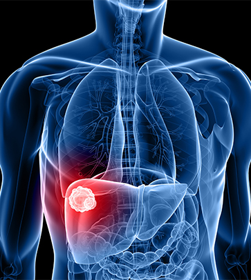
Liver cancer (hepatocellular carcinoma) is a cancer arising from the liver. It is also known as primary liver cancer or hepatoma. The liver is made up of different cell types (for example, bile ducts, blood vessels, and fat-storing cells). However, liver cells (hepatocytes) make up 80% of the liver tissue. Thus, the majority of primary liver cancers (over 90%-95%) arises from liver cells and is called hepatocellular cancer or carcinoma. 

When patients or physicians speak of liver cancer, however, they are often referring to cancer that has spread to the liver, having originated in other organs (such as the colon, stomach, pancreas, breast, and lung). More specifically, this type of liver cancer is called metastatic liver disease (cancer) or secondary liver cancer. 

This is a much more common problem around the world than primary liver cancer and frequently leads to confusion, because the term liver cancer actually can refer to either metastatic liver cancer or hepatocellular cancer.
What are the Symptoms of Liver Cancer
Most of the time liver cancer in the early stages does not cause symptoms. If symptoms are present, they may include: unexplained weight loss on-going lack of appetite fullness after a small meal a swollen liver or a mass that can be felt in the area of the liver ongoing stomach pain extending to the back and shoulder a swollen abdomen yellow-green color to the skin and eyes (jaundice) increased symptoms of illness in those who have chronic hepatitis or cirrhosis.
Categories
Medreads

Gall Bladder Cancer

Gall Bladder Cancer

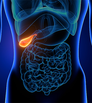
The gall bladder is a small pouch that stores and concentrates bile. Bile is a fluid that helps us to digest food. Its main function is to break down fats in food. Bile is made by the liver and stored in the gall bladder. The gall bladder is connected to the small intestine and the liver by the bile ducts.

Cause and Risk Factors

Most of the time liver cancer in the early stages does not cause symptoms. If symptoms are present, they may include:

  • Gall stones and inflammation: Gall bladder cancer is more likely to occur in people who have a history of gallstones or in people who have inflammation of the gall bladder (cholecystitis). However, most people who have gallstones or an inflamed gall bladder won’t develop gall bladder cancers.
  • Polyps: These are non-cancerous (benign) tumours of the gall bladder that increase the risk of developing gall bladder cancer.
  • Abnormal bile ducts: Gall bladder cancer is slightly more common in people who are born with (congenital) abnormalities of the bile ducts.
  • Porcelain gall bladder: People who have a condition called porcelain gall bladder, in which calcium forms in the wall of the gall bladder, also have a slightly increased risk of this type of cancer.
  • Smoking: Some evidence suggests that people who smoke cigarettes are more likely to develop gall bladder cancer.
  • Family history: People who have a close relative (parent, brother or sister) with gall bladder cancer have a slightly higher risk of developing this type of cancer.
  • Obesity: Being overweight increases your risk of developing gall bladder cancer.Early gall bladder cancer often causes no symptoms and is usually discovered unexpectedly when someone has surgery to remove gallstones. About 1 in 5 gall bladder cancers are found in this way.

Most tumours are only discovered at an advanced stage. They can cause a variety of symptoms, including sickness, high temperatures, weight loss and pain in the tummy (abdomen).

If the cancer blocks the bile duct, it may stop the flow of bile from the gall bladder into the small bowel. This causes bile to flow back into the blood and body tissues, and leads to the skin and whites of the eyes becoming yellow (known as jaundice).

The urine also becomes a dark yellow colour and stools (bowel motions) are pale. The skin may become itchy.

These symptoms may be caused by other problems, such as gallstones or infection of the gall bladder, but it’s important to get them checked by your doctor.

Categories
Medreads

Oesophageal Cancer

Oesophageal Cancer

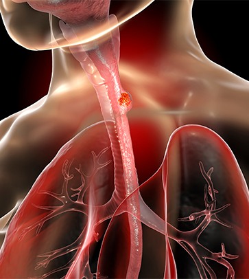
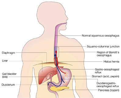
Cancer of the oesophagus is a disease in which malignant cells arise from the tissues of the oesophagus (tube leading to stomach). These eventually grow and obstruct the oesophagus, and spread to other parts of the body, such as the liver. There are two main types of oesophageal cancer: adenocarcinoma and squamous cell carcinoma. Most adenocarcinomas are now thought to arise in Barrett’s mucosa (see Barrett’s Oesophagus). The most common sign of cancer of the oesophagus is difficulty in swallowing (dysphagia). Pain in the chest may be felt when swallowing or at other times. Loss of appetite and weight loss will occur. As the oesophagus becomes obstructed, food which has not passed down the oesophagus may spill over into the lungs (aspiration), causing pneumonia.

Medical Treatment

The decision regarding treatment is complex. There are several options:

  • Surgery to remove the cancer, with intent to cure
  •  Surgery to remove the cancer, to provide palliation (relief)
  • Radiotherapy and chemotherapy (alone or combined with surgery)
  •  Photodynamic therapy (for very early cancers in selected patients)
  • Laser therapy (mainly palliative if the cancer is large)
  •  Stenting (insertion of a tube to prevent the cancer obstructing the oesophagus)

The choice of treatment depends on many factors including the size of the cancer and extent of spread, age and fitness of the patient, and the patient’s wishes. The chance of recovery (prognosis) depends on the stage of the cancer (whether it is in the oesophagus or if it has spread outside to other tissues) and the patient’s general state of health.